Copyright © 2014: Manila Retina Specialists All Rights Reserved
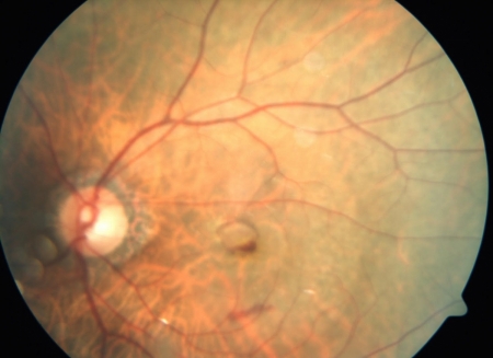
 Fig. 10 Patient sees a dark spot in center vision.
This is a condition where the part of the retina we use to read (macula) is damaged by abnormal blood vessels or death of the Retinal Pigment Epithelium (RPE), the cells which keep the macula healthy. Without the RPE, debris is not cleared as efficiently, hence the retina over this area dies or atrophies.
This may be a slow onset of blurred central vision in one or both eyes. Patient usually notices that he can see the body of the person he is talking to, but cannot see some features of his face.
Some patients present will metamorphopsia, or distortion of objects. Some may have sudden onset of dark black spot in his field of vision. This may be due to bleeding under the retina.
There are two types, wet and dry. The wet type is treated with Anti-VEGF injections in combination with multivitamins, zinc and lutein. The dry type is treated with multivitamins alone, but there are various research on the use of stem cells for this type of macular degeneration.
 Fig 11. Wet type of macular degenerationIn Asian population like ours, Idiopathic Polypoidal Choroidal Vasculopathy (IPCV) is a common disorder which simulates the symptoms of wet type of macular degeneration. OCT (Optical Coherence Tomography) helps us diagnose and monitor this condition. In some cases, your doctor may order Fluorescein/ICG Angiography to help us determine if a polyp is the cause of the bleeding.
In most cases, monotherapy with ant-VEGF drugs is enough. In certain situations, depending on the location of the polyp, focal laser, or PDT (Photodynamic Therapy) may be used to destroy the polyp, in addition to anti-VEGF (Avastin, Lucentis, or Eylea) treatment.
