Copyright © 2014: Manila Retina Specialists All Rights Reserved
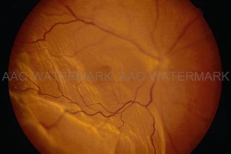
Retinal Detachment is a condition where there is separation of the layers of the retina, usually due to a tear, or membranes caused by diabetes, vein occlusions or inflammatory conditions (uveitis). It is usually heralded by the sudden onset of floaters, flashes, or a shadow or curtain in your field of vision.
 Fig 5 Corrugated appearance of retina in detachment cases. Fig 5 Corrugated appearance of retina in detachment cases. | photo courtesy of American Academy of Ophthalmology (AAO) |
Signs & symptoms
Floater & Flashes of Light- the eye ball is filled with thick vitreous gel. As we age the gel like becomes less viscous. As the vitreous becomes liquefied and pulls away from the retina, it becomes somewhat condensed and stringy and forms strands which may be perceived as floaters.
As the eyeball moves, the liquefied vitreous moves around the vitreous cavity. Because of this movement, the vitreous begins to pull the retina. With time, the vitreous can pull free and separate from the retina and optic nerve and this is called POSTERIOR VITREOUS DETACHMENT (PVD). When a person develops PVD, flashes of light or dark spots in the vision may occur. Pictures courtesy of AAO.
Treatment
Depending on what your case is, the doctor may recommend Pars Plana Vitrectomy, Scleral Buckling, Pneumatic Retinopexy, or combination treatment. Most can be done in an outpatient setting, but if procedure will take a long time, we may recommend hospital admission. Your doctor will recommend the best option for you.
To keep the retina in place, your Retina Surgeon may put special long acting gases, or medical grade silicone oil. Intraocular gases may need air travel restrictions, and positioning, for a certain period of time.
A Retina Specialist is an Ophthalmologist who, in addition to a 3 year residency program, trains for another year or two to perform Retina and/or Vitreous Surgery.
